
Welcome to my rhabdomyolysis page. This is actually a copy of an assignment I did for my fourth year medicine neuropathology course back in 1997. Whilst doing this assignment I found that there was not much information about this topic on the web, so I thought it might be useful to put online. So here it is, everything I was able to find about rhabdomyolysis. I hope you find it helpful. If you would like more information please speak to your doctor. I am not a specialist in this area and would not be of any help beyond what I have put in this web page, which was done quite a while back, and has not been revisited since. Nevertheless, I hope this information is of value.
OUTLINE
 LINKS
LINKS
 Dr Baggas' Surgery
Baggas' World
INTRODUCTION
Rhabdomyolysis is a common disorder which may result from a large variety
of diseases, trauma, or toxic insults to skeletal muscle. It may be defined
as a clinical and biochemical syndrome resulting from an injury which damages
the integrity of the sarcolemma of skeletal muscle, leading to the release
of potentially toxic muscle cell components into the circulation.(1,2,3)
This may result in potential life-threatening complications including myoglobinuric
acute renal failure, hyperkalaemia and cardiac arrest, disseminated intravascular
coagulation, and more locally, compartment syndrome.
BIOCHEMISTRY
The primary diagnostic indicator of rhabdomyolysis is an elevated
serum creatine phosphokinase (CK) to at least five times the normal value.(2)
This elevation is generally to such a degree that myocardial infarction
and other causes of a raised CK are excluded. Additionally, the CK-MM isoenzyme
predominates in rhabdomyolysis, comprising at least 98% of the total value.(4)
The other important finding frequently seen in rhabdomyolysis is myoglobinuria.
Myoglobin, a haem protein which functions as an oxygen store in type 1
skeletal muscle fibres, normally has a rapid renal clearance which maintains
a low plasma level up to a certain serum concentration.(5) As myoglobin
is released into the circulation from necrotic muscle cells it first becomes
detectable in the urine at serum concentrations ranging from 300ng/ml to
2 g/ml and produces visible pigmenturia (classically a "coca-cola"
coloured urine) at concentrations exceeding 250 g/ml.(6) This discolouration
is caused by myoglobin plus metmyoglobin in the urine.(7) Biochemical tests
for pigmenturia are strongly suggestive of myoglobinuria in the absence
of haemoglobinaemia and haematuria.(7) Other important biochemical findings
in rhabdomyolysis include hyperkalemia, hypocalcaemia, hyperphosphataemia,
hyperuricaemia, and raised levels of other muscle enzymes including lactate
dehydrogenase, aldolase, aminotransferases, and carbonic anhydrase III
(which is a very specific marker for skeletal muscle injury).(2) Metabolic
acidosis may result from release of phosphate, sulphate, uric acid, and
lactic acid from the muscle cell.(1)
AETIOLOGY
The causes of rhabdomyolysis can be broadly divided into hereditary (table
1) and acquired (table 2) groups. The hereditary
causes consist primarily of enzyme defects causing disorders of carbohydrate
metabolism(8), mitochondrial lipid metabolism(8), and other inherited disorders
such as malignant hyperthermia (8,9) and neuroleptic malignant syndrome(10).
Table 1 : Inherited causes of rhabdomyolysis.
(from Poels and Gabreels
(1993) Clin Neurol Neurosurg 95 : 175-192.
Deficiencies of glyco(geno)lytic enzymes
myophosphorylase (McArdle's disease)
phosphorylase kinase
phosphofructokinase (Tarui's disease)
phosphoglycerate mutase
phosphoglycerate kinase
lactate dehydrogenase
Abnormal Lipid Metabolism
carnitine palmitoyltranferase deficiency I and II
carnitine deficiency
Other genetic disorders
idiopathic rhabdomyolysis
myoadenylate deaminase deficiency
malignant hyperthermia
neuroleptic malignant syndrome
Acquired causes may be divided into traumatic, ischaemic, metabolic, infectious,
inflammatory, and toxic groups(table 3) (11), as
well as exercise and heat related causes.
Table 2 : Acquired causes of rhabdomyolysis.
(from Poels and Gabreels
(1993) Clin Neurol Neurosurg 95 : 175-192.
Toxic
alcohol
drugs and toxins (see Table 3)
Excessive muscle exercise
sports and military training
status epilepticus
status asthmaticus
convulsions
prolonged myoclonus, acute dystonia
Direct muscle injury
crush
burning, freezing
electric shock, lightning stroke
Ischemic injury
compression
vascular occlusion
sickle cell trait
Metabolic disorders
diabetic ketoacidosis
nonketotic hyperosmolar coma
hypothyroidism
hypophosphatemia
hyponatremia
hypokalemia
Infections
bacterial
viral
Heat-related syndromes
toxic shock syndrome
heat stroke
Inflammatory myopathies
polymyositis
dermatomyositis
Others
anticholinergic syndrome
withdrawal of L-Dopa
Table 3 : Drugs and toxins known to cause rhabdomyolysis.
(11)
Drug-induced coma, seizures,dyskinesia Other drugs
Barbiturates Amphetamines
Heroin Phenmetrazine
Methadone Phencyclidine
Glutethimide Phenylpropanolamine
Chlorpromazine Morphine
Diazepam Dihydrocodeine
Rohypnol LSD
Lithium Salicylates
Amoxapine Clofibrate/Bezafibrate
Phenelzine` Epsilon-aminocaproic acid
Phenformin/fenfluramine Isoniazid
Meprobamate Loxapine
Antihistamines/paracetamol Theophyllin
Oxprenolol Pentamidine
Ethanol Vasopressin
Post-anaesthetic Toxins
Suxamethonium Ethanol
Malignant hyperpyrexia Isopropyl alcohol
Carbon monoxide
Neuroleptic malignant syndrome Mercuric chloride
Haloperidol Ethylene glycol
Stelaziine Copper sulphate
Fluphenazine Zinc phosphide
Other neuropleptics Strychnine
Metaldehyde
Chloralose
Hypokalaemia Paraphenylenediamine
Diuretics Toluene (paint sniffing)
Carbenoxolone Gasoline sniffing
Amphotericin B Lindane/benzene
Liquorice Snake bite
Hornet/wasp sting
Brown spider bite
Haff disease
Quail ingestion
PATHOGENESIS
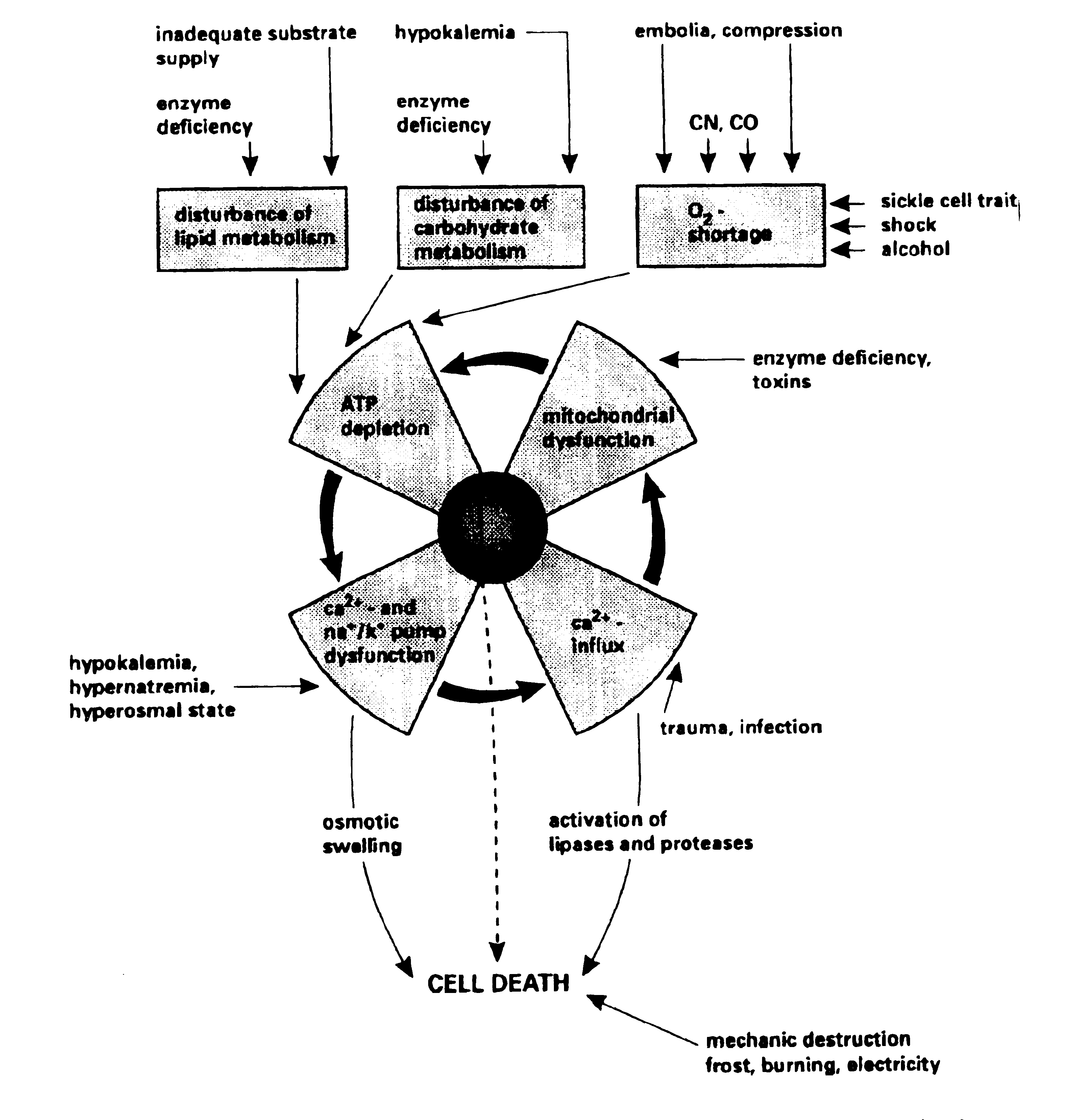
Although the causes of rhabdomyolysis are so diverse, the pathogenesis
appears to follow a final common pathway, ultimately leading to muscle
necrosis and release of muscle components into the circulation. Whatever
the injurious process, the end result is an increased cellular permeability
to sodium ions due to either plasma membrane disruption or reduced cellular
energy (ATP) production.(1) Accumulation of sodium in the cytoplasm leads
to an increase in intracellular calcium concentration (which is normally
very low relative to the extracellular concentration).(2) This accumulation
of calcium is due both to direct injury to the cell and to increased activity
of an Na+/Ca2+ exchanger protein which brings more calcium into the cell
as it attempts to remove the excess sodium. Depletion of ATP also contributes
directly to calcium accumulation due to a reduction in the activity of
the Ca2+ ATPase which normally acts to pump calcium out of the cell and
sequester it in the sarcoplasmic reticulum.(3)
Therefore, the common pathogenetic feature of all disease processes causing
rhabdomyolysis is an acute rise in the cytosolic and mitochondrial calcium
concentration in affected muscle cells, which sets off a chain of events
that ultimately results in muscle cell necrosis. This includes activation
of degradative enzymes such as phospholipase A2 (PLA) and neutral proteases,
leading to membrane phospholipid and myofibril damage.(3) Jackson et al.
(12) suggest that the most significant of these is activation of PLA, and
that most of the membrane and mitochondrial damage in rhabdomyolysis can
be attributed to this. PLA mediated attack on mitochondrial and sarcolemmal
membrane phospholipids leads to the formation of lysophospholipids and
free fatty acids. These further potentiate the injury by causing direct
membrane damage themselves and through alterations in ionic transport,
which results in further influx of sodium and calcium.(3) Thus the reaction
becomes self-perpetuating. Depletion of ATP and mitochondrial damage may
be the primary event which sets off this cascade (as in most hereditary
causes of rhabdomyolysis and exertional rhabdomyolysis) or it may occur
secondarily to the rise in calcium concentration. Either way, mitochondrial
damage and depletion of ATP contributes to the pathogenesis via the following
:
(1) Failure of Ca2+ ATPase leading to failure of calcium sequestration
and reduced efflux of calcium from the cell.
(2) Failure of Na+/K+ ATPase leading to increased intracellular sodium
and increased Na+-Ca2+ exchange, further contributing to the increased
intracellular calcium.2
(3) Generation of toxic oxygen free radicals such as superoxide causes
further cellular damage.(3)
A simple schematic representation of these processes is shown in Figure
1 . Ultimately, the combination of all of these processes is a self-sustaining
reaction which results in muscle cell lysis (figure2) and release of intracellular
components into the extracellular fluid and circulation.(3) Locally, accumulation
of these products may result in microvascular damage, capillary leak and
increased intracompartmental pressures, and reduced tissue perfusion and
ischaemia, which may further potentiate the muscle damage.
As has already been stated, there are many different causes of rhabdomyolysis,
and although the final reaction is fairly stereotyped, the mechanism by
which this reaction is triggered is quite variable. I will now discuss
some of the specific hereditary and acquired causes of rhabdomyolysis in
more detail.
HEREDITARY CAUSES OF RHABDOMYOLYSIS
Disorders of Muscle Carbohydrate Metabolism
The first genetic disease described which causes rhabdomyolysis is McArdle's
disease (myophosphorylase deficiency), an autosomal recessive condition
in which there is selective necrosis of type 2 muscle fibres.(8) These
fibres are more dependent on glyocolysis for generation of ATP and therefore
will be more sensitive to an enzyme defect which prevents the formation
of glucose from glycogen. Hence it is ATP depletion which is responsible
for rhabdomyolysis in this disease. Muscle pain and rhabdomyolysis are
induced by vigorous exercise,and relieved by rest in this disease, consequently
patients can adjust their life styles to prevent symptoms by avoiding vigorous
exercise which requires activation of type 2 fibres. Other inherited diseases
affecting the glycolytic/ glycogenolytic pathways include
phosphofructokinase deficiency (Tarui's disease), and phosphoglycerate
mutase deficiency.(8)
Carnitine Palmitoyltransferase Deficiency
Where the disorders of carbohydrate metabolism affect primarily anaerobic
type 2 muscle fibres, diseases of lipid metabolism such as Carnitine palmitoyltransferase
deficiency (CPD), have a greater effect on aerobic type 1 fibres
which depend on the oxidation of long chain fatty acids to produce energy.
CPD, an autosomal recessive disorder, has been shown to be the most common
hereditary disease causing rhabdomyolysis.(3) In this disease muscle pain
and rhabdomyolysis develop after prolonged exercise with inadequate nutrient
intake, not in the initial phase as in the glycogen storage disorders.
Treatment of this disease involves frequent high carbohydrate meals and
avoidance of prolonged exercise.(8)
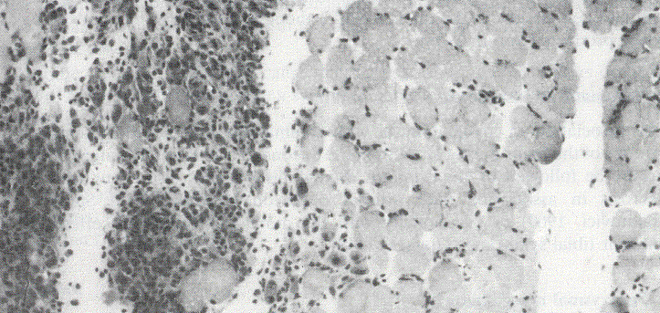
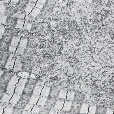
Malignant Hyperthermia
Another genetic disease which may result in rhabdomyolysis is malignant
hyperthermia (MH) (Figure 3). In this disease, episodes of hyperthermia and rhabdomyolysis
are triggered by exposure to volatile anaesthetics such as halothane, or
succinylcholine, a depolarising muscle relaxant.(9) MH appears to be an
autosomal dominant condition with variable penetrance(7), and may involve
a defect in the ryanodine receptor of the calcium release channel of the
sarcoplasmic reticulum.(7) These patients have higher than normal resting
sarcoplasmic calcium concentrations, and exposure to the above agents may
trigger further uncontrolled calcium release, leading to excessive muscle
contraction, hyperthermia, and rhabdomyolysis.(8) The diagnosis of MH susceptibility
can be made only by muscle biopsy and a positive in vitro response to provocative
agents such as halothane, succinylcholine, and caffeine. This in vitro
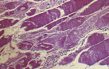
response shows a patchy, moth-eaten appearance of type 1 fibres(13) (Figure 4). Type
1 fibres are predominantly affected in MH due to their lower capacity for
anaerobic metabolism, and therefore more rapid ATP depletion in the hypermetabolic
state of MH.
Neuroleptic Malignant Syndrome
A similar disorder is the Neuroleptic Malignant Syndrome (NMS), in which
there is a gradual development of hyperthermia, muscle rigidity, fluctuating
consciousness, and autonomic instability.(10) Rhabdomyolysis and myoglobinuria
may result. Drugs which can cause NMS include phenothiazines, butyrophenones,
and other antipsychotics and antidepressants. It is believed that the underlying
defect in NMS may be a central or presynaptic one, in contrast to the peripheral
defect in MH. (10)
ACQUIRED CAUSES OF RHABDOMYOLYSIS
There are also many non-hereditary causes of rhabdomyolysis, which are
much more common than the hereditary causes.
Exertional Rhabdomyolysis
Exertional rhabdomyolysis and heat stroke are probably the most common
causes of severe rhabdomyolysis. This occurs most commonly in untrained
people undertaking vigorous exercise in hot, humid weather.(3) The pathogenesis
of rhabdomyolysis in these cases appears to be due to a combination of
mechanical and thermal muscle injury and ATP depletion, both of which ultimately
lead to calcium accumulation. Excess muscle activity may also lead to rhabdomyolysis
in conditions such as generalised seizures, status epilepticus, status
asthmaticus, myoclonus, and severe dystonia. (2)
Crush Injury and Trauma
In crush injury and other forms of trauma, rhabdomyolysis is generally
due to direct muscle injury and ischaemia. However, in addition to this,
in the crush injury, reperfusion after prolonged ischaemia is also believed
to play a significant role in muscle damage.(14) This is believed to be
mediated by the formation of oxygen free radicals, the action of granulocytes,
and increased calcium uptake after ischaemia (which is due to exchange
of calcium for excess intracellular sodium which has accumulated during
the ischaemic period).
Alcoholism
Alcoholism is another common cause of rhabdomyolysis. This may be secondary
to to alcohol related trauma, seizures, or coma, or may be due to a direct
toxic effect of ethanol on skeletal muscle, resulting in both a chronic
myopathy, and acute rhabdomyolysis.(3)It is believed that ethanol causes
direct sarcolemmal injury, leading to increased sodium permeability, and
subsequent accumulation of calcium.1 Hypophosphataemia may be an important
precipitant of rhabdomyolysis in alcoholics, since the ability of muscle
cells to produce ATP would be reduced. (4)
Drugs and Toxins
A large range of drugs and toxins have been seen to cause rhabdomyolysis.
Many of these are listed in Table 3. The mechanisms of muscle damage in
these instances are diverse.
Some drugs appear to have a direct toxic action on skeletal muscle when
given systemically. These include cholesterol lowering drugs (clofibrate,
gemfibrozil, HMG CoA reductase inhibitors), emetine (ipecac), zidovudine
(AZT), vincristine, and epsilon-aminocaproic acid(Figure 5).(15,11)
An immunological mechanism may be responsible for the myositis seen in
patients treated seen in patients treated with D-penicillamine, L-tryptophan,
and rarely in other drugs including procainamide, cimetidine, phenytoin,
and levodopa.(11)
Amphotericin B, carbenolexone, liquorice, laxatives, and diuretics may
cause rhabdomyolysis secondary to sever hypokalaemia.(11)
Another mechanism by which drugs may cause rhabdomyolysis is by excessive
neuromuscular stimulation. These drugs include phencyclidine (PCP), and
acetylcholinesterase inhibitors.(11)
Drugs such as heroin and barbiturates may contribute to rhabdomyolysis
via coma and muscle compression following overdose.(2)
In addition to the range of pharmacologic agents which cause rhabdomyolysis,
it can also be caused by the venoms of a number of snakes, spiders, and
wasps.(11) Microbial toxins such as the a-toxin of Clostridium perfringens
(gas gangrene), can also cause rhabdomyolysis, as can excessive consumption
of quail. (11)
CLINICAL FEATURES
The clinical features of rhabdomyolysis are quite variable, no doubt due
to the large range of causes of this condition. Broadly, they can be divided
into the following2 :
(1) Muscular signs and symptoms
(2) General internal disturbances
(3) Complications
Muscular signs and symptoms
These include pain, weakness, tenderness, and contractures. This may involve
specific groups of muscles or may be generalised. Most frequently the involved
muscle groups are the calves and lower back, however a significant proportion
may show no signs of muscle injury at all.(16) Sometimes haemorrhagic discolouration
of the overlying skin may be seen. Typically the muscle disorder is self-limiting
and resolves within days to weeks, due to the regenerative capacity of
muscle.
General internal disturbances
These include malaise, fever, tachycardia, nausea, and vomiting. Hyperuricaemia
may lead to encephalopathy with depression of respiration with hypoxia
and respiratory acidosis. (2)
COMPLICATIONS
The complications of rhabdomyolysis are due to the local effects of muscle
injury, and the systemic effects of released muscle components. These include
:
(1) Hypovolaemia - due to haemorrhage, and influx of fluid into
necrotic muscle. 4-11 litres of normal saline may be required to maintain
cardiac and urine output. (2,16)
(2) Cardiac arrest and arrhythmias - Hyperkalaemia can precipitate
severe arrhythmias and cardiac arrest. This toxicity is potentiated by
the hypocalcaemia resulting from calcium deposition in necrotic muscle.
Therapy often involves the use of ion exchange resins.
(3) Compartment Syndrome - (Figure 6)in acute rhabdomyolysis muscle swelling
within a tight fascial compartment can lead to compression of vessels and
nerves. This can lead to nerve damage and muscle ischaemia due to reduced
capillary flow. Ischaemia will result in further oedema which prolongs
the cycle. Prolonged ischaemia and infarction of muscle tissue can result
in replacement of muscle by inelastic fibrous tissue and severe contractures
(Volkmann's contracture).(17,2) The treatment of suspected compartment
syndrome is urgent decompression by open fasciotomy.
(4) Disseminated intravascular coagulation - this is an almost universal
finding in patients with rhabdomyolysis (18) and is probably due to activation
of the clotting cascade by released muscle components. Fortunately, in
most cases, the diagnosis of DIC is made purely by laboratory abnormalities
rather than overt clinical bleeding or thrombosis.(16)
(5) Acute Renal Failure - this is probably the
most significant and most feared complication of rhabdomyolysis, and is
said to occur in about 30% of patients.(16) Conversely, rhabdomyolysis
has been said to be a factor in 8% of cases of acute renal failure2 so
this is by no means an uncommon condition. The mechanisms of myoglobinuric
acute renal failure have been comprehensively explored by Zager (1996)
(3) and include the following :
(1) Renal vasoconstriction/hypoperfusion - due to hypovolaemia and
haem- protein induce renal tubular ATP depletion
(2) Haem protein cast formation - precipitation of pigment casts
in distal tubules may contribute to acute tubular necrosis, especially
in aciduria
(3) Ischaemic tubular injury - independent of haemodynamic influences,
haem protein can potentiate proximal tubular ischaemic damage
(4) Haem iron induced oxidant stress - intratubular release of haem
iron catalyses formation of toxic oxygen free radicals
Prevention of myoglobinuric ARF involves maintenance of circulating blood
volume by adequate fluid replacement of up to 11 litres of normal saline.
(2) Administration of frusemide and/or mannitol is used to maintain a diuresis
and enhance haem protein elimination. Alkalinization of the urine by the
addition of sodium bicarbonate to the intravenous fluids has been suggested
(since acidic urine favours myoglobin nephrotoxicity) however this is controversial
since bicarbonate may aggravate existing hypocalcaemia. (2,3)
CONCLUSIONS
Rhabdomyolysis is a common condition which complicates a a variety of genetic
and acquired diseases. It is characterised by muscle cell necrosis and
release of muscle cell components into the circulation, most notably creatine
phosphokinase (CK) and myoglobin. The primary mechanism through which muscle
damage occurs in rhabdomyolysis is sarcoplasmic calcium overload leading
to activation of degradative enzymes. This may occur secondary to a number
of processes including ATP depletion and increased intracellular sodium
concentration, and direct sarcolemmal injury. The complications of rhabdomyolysis
can be potentially life threatening, and include cardiac arrest and myoglobinuric
acute renal failure. Prompt action must be taken to prevent these complications
in a patient with rhabdomyolysis, most importantly aggressive intravenous
volume replacement.
REFERENCES
1. Knochel, J.P. (1993) Mechanisms of rhabdomyolysis. Current Opinion in Rheumatology 5: 725-731.
2. Poels, P.J.E and Gabreëls, F.J.M. (1993) Rhabdomyolysis : a review of the literature. Clin Neurol & Neurosurg 95: 175-192.
3. Zager, R.A. (1996) Rhabdomyolysis and myohemoglobinuric acute renal failure. Kidney International 49 : 314-326.
4. Knochel, J.P. (1992) Hypophosphataemia and rhabdomyolysis. JAMA 92: 455-457.
5. Dayer-Berenson, L. (1994) Rhabdomyolysis : A comprehensive guide. ANNA Journal 21(1): 15-18.
6. Penn, A.S. (1986) Myoglobinuria. In: Engel, A.G, and Banker, B.Q. (Eds) Myology, Vol 2. New York : McGraw-Hill, 1785-1805.
7. Moxley, R.T. (1994) Metabolic and endocrine myopathies. In: Walton, J., Karpati, G., and Hilton-Jones, D. (Eds) Disorders of Voluntary Muscle (6th ed). New York : Churchill Livingstone, 647-716.
8. Brumback, R.A., Feeback, D.L., and Leech, R.W. (1992) Rhabdomyolysis in childhood. Paediatric Neurology 39(4) : 821-858.
9. Gronert, G.A. (1986) Malignant Hyperthermia. In: Engel, A.G, and Banker, B.Q. (Eds) Myology, Vol 2. New York : McGraw-Hill, 1763-1784.
10. Guzé, B.H. and Baxter, L.R. (1985) Neuroleptic Malignant Syndrome. NEJM 313(3): 163-166.
11. Kakulas, B.A. and Mastaglia, F.L. (1992) Drug-induced, toxic and nutritional myopathies. In: Mastaglia, F.L. and Walton, J. (Eds) Skeletal Muscle Pathology (2nd ed). New York : Churchill Livingstone, 511-540.
12. Jackson, M.J., Jones, D.A., and Edwards, R.H.T. (1984) Experimental skeletal muscle damage : the nature of the calcium activated degenerative processes. Eur J Clin Invest 14: 369-374.
13. Anderson, J.R. (1985) Atlas of Skeletal Muscle Pathology. Lancaster : MTP Press.
14. Odeh, M. (1991) The role of reperfusion-induced injury in the pathogenesis of the crush syndrome. NEJM 324(20) : 1417-1422.
15. Argov, Z. and Mastaglia, F.L. (1994) Drug-induced neuromuscular disorders in man. In: Walton, J., Karpati, G., and Hilton-Jones, D. (Eds) Disorders of Voluntary Muscle (6th ed). New York : Churchill Livingstone, 989-1029.
16. Saad, E.B. (1997) Rhabomyolysis and Myoglobinuria. (internet reference : http://www.medstudents.com.br/terin/terin3.htm)
17. Apley, A.G. and Solomon, L. (1994) Concise System of Orthopaedics and Fractures. Oxford : Butterworth-Heinemann.
18. Knochel, J.P. (1990) Catastrophic medical events with exhaustive exercise : "White collar rhabdomyolysis". Kidney International 38: 709-719.
